Historically, the types of kava products (chemotype) have been characterized by their relative abundance of individual kavalactones [ 92 ]. Kava products of different chemotypes have been proposed to possess varied benefits and risks, because of different composition profiles of kavalactones and flavokavains [ 93 ]. Other than in the format of a drink, kava has also been commercialized in the form of capsules or tinctures as dietary supplements. Because of these variables, currently available kava products could be very diverse due to their difference in format, kavalactone abundance and profiles, and the content of other ingredients, such as flavokavains A and B [ 94 96 ]. Not surprisingly, various benefits and risks could potentially be introduced due to these chemical composition variations. Commercial kava products thus should be rigorously standardized with accurate content information for human use, particularly in the case of potential chronic use, such as its potential use in primary lung carcinogenesis prevention [ 95 ].
Despite their high structural similarity, these natural kavalactones have distinct pharmacokinetic [ 87 ] and pharmacodynamic properties [ 88 89 ]. It is therefore possible that these kavalactones may be complementary to each other and none of them individually will be able to fully recapitulate the holistic beneficial properties of kava. Other than kavalactones, a class of chalcone based compounds have been detected in kava products and heavily investigated, named flavokavains A, B, and C ( Figure 2 ) [ 90 ]. These flavokavains have not been reported to contribute to kava’s relaxing property. A number of putative targets have been reported for kavalactones, including voltage-gated sodium and calcium ion channels, gamma-aminobutyric acid (GABA) type A receptors, and monoamine oxidase B (MAO-B), with detailed information in our previous review [ 91 ].
In addition, cancer incidence rates were lower in males relative to females in South Pacific nations with higher kava consumption, which is the opposite of global trends, as previously mentioned [ 97 ]. Given that traditional kava is dominantly consumed by males, the lower cancer incidence among males versus females in nations with high kava consumption also supports kava’s potential to prevent cancer. In this report, cancer incidence includes all types of cancers. It is possible that kava may have differential effects among different cancer types, which has not yet been rigorously investigated. A limited number of potential confounding variables other than kava were briefly analyzed as well, such as smoking rate, which was found to be comparable among those nations and may not contribute to the observed cancer incidence differences [ 98 ]. Several other epidemiological data also indicate lower cancer incidence among males in comparison to females in the South Pacific [ 99 100 ], which is again opposite to the global trend [ 1 ], further substantiating kava’s potential in reducing human cancer risk.
4.3. Kava’s Potential in Cancer Risk Reduction in Animal Models, Responsible Ingredients, and Mechanisms
54,89,101,102,103,104,105,106,107,108,109,110, Stimulated by these interesting human epidemiological data, kava’s potential to reduce cancer risk has been evaluated during the past two decades using various chemical-induced or transgenic animal models, including lung, prostate, colon, and bladder tumorigenesis [ 53 111 ]. In these animal models, tumorigenesis was induced by genetic mutations or different chemical carcinogens via different administration routes. Kava, via gavage or in the form of diet, has also been administered via different regimens, either during or after carcinogen exposure in the chemical carcinogenesis model. The positive results of kava to prevent tumorigenesis in all of these animal models strongly suggest that kava may reduce human cancer risk, likely via different mechanisms. In fact, different kavalactones have been identified as the active ingredients in some of these carcinogenesis models. For instance, dihydromethysticin has been identified as one active compound that can effectively suppress NNK-induced lung carcinogenesis in A/J mice [ 89 ] while kavain was recently identified to prevent bladder carcinogenesis induced by hydroxy butyl(butyl) nitrosamine (OH-BBN) in mice [ 111 ]. Although kavain has not been evaluated for its potential against NNK-induced lung carcinogenesis, it is less likely to be as effective as dihydromethysticin in this model based on its lack of efficacy in reducing NNK-induced DNA damage in target lung tissue [ 89 ]. These results also argue that maybe none of the single-chemical entities in kava are capable of fully recapitulating the holistic benefits of kava in cancer risk reduction, upon which the human epidemiological data are built. This is particularly important for human translation. Kava, a natural blend of kavalactones with historical human exposure and epidemiological support, may be the ideal candidate instead of any single chemical from kava as long as the kava product has rigorous quality control and quality assurance.
102, With respect to its potential in preventing lung carcinogenesis, kava was first evaluated against lung carcinogenesis induced by eight oral dosages of NNK and BaP in A/J mice [ 53 ]. Kava was supplemented in the diet with three different treatment regimens, covering only the carcinogen exposure period (mimicking current smokers), covering the postcarcinogen exposure period (mimicking former smokers), and covering the whole experimental period. In all of these treatment regimens, kava significantly reduced the number of lung tumors, indicating kava’s potential to reduce lung cancer risk among both current and former smokers [ 53 ]. Preliminary mechanistic investigation suggests that kava inhibited the activation of nuclear factor kappa B (NF-κB) [ 53 ]. Given that chronic lung inflammation is a well-established risk factor for lung cancer, kava may prevent lung carcinogenesis in this animal model at least in part by suppressing tobacco-induced lung inflammation. Chalcone-based flavokavains ( Figure 2 ) in kava were initially hypothesized as the responsible active ingredients since many chalcone-based compounds have been reported with cancer preventive potential in various animal models [ 112 ]. Our data later rejected this hypothesis as flavokavains from kava, at several dosages, failed to capture the preventive efficacy of kava in this animal model [ 104 ]. Additional medicinal chemistry efforts from our lab were able to develop analogs of the flavokavains with a wide range of bioactivity in cell models, but none of them were able to block lung carcinogenesis in this animal model and some compounds showed significant toxicity (unpublished data). The traditional approach for active ingredient identification, fractionation and biological evaluation, was adopted to search for the active chemicals [ 54 ]. The fraction enriched with kavalactones was able to recapitulate the preventive efficacy of kava in a two-dose NNK-induced lung carcinogenesis A/J mouse model and dihydromethysticin was identified as an active compound [ 54 ]. Dihydrokavain was demonstrated completely inactive in this animal model, which later was used as a control compound for mechanistic elucidation. Based on their distinct effects in reducing NNK-induced DNA damage, methysticin is likely active as well while kavain would not in this animal model [ 54 ]. Extensive structure–activity relationship studies have been performed on dihydromethysticin to characterize the functional groups important for its lung cancer preventive activity [ 101 108 ] but to date none of the synthetic compounds were able to outperform the natural dihydromethysticin except the unnatural enantiomer of dihydromethysticin. It should be noted that the dose range of kava and natural dihydromethysticin, with effective lung cancer prevention in these animal studies, was comparable to the levels of traditional kava consumption in humans. Thus, in alignment with the epidemiologic data, kava may be potent enough to reduce human lung cancer risk in its natural format.
106, In the two-dose NNK-induced lung tumorigenesis model, one mechanism of kava to prevent lung carcinogenesis is to enhance NNK detoxification and thus reduce NNK-induced DNA damage [ 106 107 ]. The enhanced NNK detoxification is likely mediated via the transcriptional upregulation of UDP-Glucuronosyltransferase (UGT) enzymes, resulting in increased glucuronidation and urinary excretion of NNAL [ 102 107 ], which may offer the potential of precision prevention given the genetic variations in UGTs in humans. At the same time, we observed that kava and dihydromethysticin may prevent lung carcinogenesis via DNA damage independent mechanisms in this animal model. Using dihydromethysticin as the example, complete tumor blockage was achieved with an incomplete protection against DNA damage (i.e., 75–88% reductions, Figure 3 ). Furthermore, dihydromethysticin given 40 h before NNK exposure reduced tumor multiplicity by 52% with little DNA damage reduction, while dihydromethysticin given concurrent with NNK reduced tumor multiplicity by only 50% although DNA damage was reduced by 63% [ 102 ]. Therefore, dihydromethysticin achieves complete prevention against NNK-induced lung carcinogenesis likely via both DNA damage-driven and -independent mechanisms [ 102 ]. The DNA damage-independent carcinogenic mechanism of NNK, however, had not been rigorously elucidated in this animal model until our recent study [ 44 ].
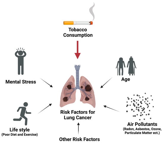
113,114,115,116,117,118,119,120,121, At the same time, chronic lung inflammation is a well-established risk factor for lung carcinogenesis. Several kavalactones have demonstrated anti-inflammatory activities in vivo [ 85 122 ]. For instance, kavain inhibits lipopolysaccharide (LPS)-induced collagen antibody induced arthritis in mice [ 120 ]. Desmethoxyyangonin inhibits LPS-induced inflammation and LPS/D-galactosamine-induced hepatitis in mice [ 113 ]. Therefore, kava may be able to reduce lung cancer risk partly through its anti-inflammatory activities. However, the two-dose NNK-induced lung tumorigenesis animal model, as discussed above, does not appear to recapitulate the chronic inflammatory nature of lung cancer risk in humans. Future work is needed to characterize the anti-inflammatory contribution of kava to reduce lung cancer risk via clinically more relevant animal models. Kava may also reduce lung cancer risk through its relaxing property if chronic mental stress is a valid risk for lung cancer. Thus, the potential contribution of stress reduction to kava’s lung cancer risk also requires future investigation. Indeed, kava revealed the potential to reduce tobacco use and tobacco dependence among smokers in a pilot clinical trial [ 123 ], which may be mediated through its relaxing properties as reflected by the reduction in the plasma levels of cortisol [ 123 ]. In summary, kava may reduce lung cancer risk via multiple mechanisms, namely reducing tobacco use and dependence, enhancing tobacco carcinogen detoxification and thus reducing DNA damage, suppressing tobacco smoke-induced lung inflammation, and promoting relaxation. How to holistically evaluate these potential benefits in a physiologically relevant animal model remains to be a major challenge.
p < 0.05). Similarly, genes significantly modified by dihydromethysticin relative to NNK were identified (1886 genes, p < 0.05). A total of 984 genes were found in common in both comparisons ( p = 5.27 × 10−225). Importantly, 89.3% of them were modified by NNK and DHM in opposite directions. These results indicate that dihydromethysticin counteracts the signaling processes induced by NNK. These genes were subjected to the Ingenuity Pathway Analysis (IPA). Protein Kinase A (PKA) was predicted as one of the top signaling pathways activated by NNK but suppressed by dihydromethysticin [126,129,130,131,134,135,137,138, With respect to the underlying molecular signaling for stress reduction and inflammation suppression, we found that NNK in tobacco smoke and its metabolite NNAL may function as β-adrenergic receptor agonists and modulate the PKA/LKB1/CREB/COX-2 pathway in A/J mouse lungs; kava and dihydromethysticin effectively suppressed the effects of NNAL on this pathway [ 44 ]. Specifically, we performed an RNA seq analysis of the A/J mouse lung tissues from control, NNK, and NNK + dihydromethysticin, respectively. Genes significantly modified by NNK relative to control were identified (3282 genes,< 0.05). Similarly, genes significantly modified by dihydromethysticin relative to NNK were identified (1886 genes,< 0.05). A total of 984 genes were found in common in both comparisons (= 5.27 × 10). Importantly, 89.3% of them were modified by NNK and DHM in opposite directions. These results indicate that dihydromethysticin counteracts the signaling processes induced by NNK. These genes were subjected to the Ingenuity Pathway Analysis (IPA). Protein Kinase A (PKA) was predicted as one of the top signaling pathways activated by NNK but suppressed by dihydromethysticin [ 44 ]. The classical PKA pathway has been well characterized: stress hormones, such as norepinephrine, bind to and activate β-adrenergic receptor (β-AR). This activates adenylyl cyclase for cAMP production. cAMP then binds to the regulatory subunit of PKA, consequently releasing and activating PRKACA (the catalytic subunit). Activated PRKACA phosphorylates CREB and induces CREB-mediated transcription, which results in the up-regulation of COX-2 that may contribute to tobacco smoke-induced lung inflammation. NNK indeed has been reported by Schuller et al. as a potent agonist for β-AR, through which it can promote NSCLC proliferation [ 124 ]. Activated PRKACA also phosphorylates LKB1, rendering LKB1 loss of its tumor suppressive function [ 80 ]. This signaling pathway was further confirmed via multiple cell models using a tobacco carcinogen metabolite, NNAL, at physiologically relevant concentrations [ 44 80 ]. Norepinephrine, the stress hormone, appears to be able to modulate the same signaling pathway (unpublished results), which may be the underlying mechanism of stress as a risk factor to primary lung carcinogenesis. In addition, these signaling events have been well-documented to contribute to cancer development and progression, including lung cancer. First, PKA activation alone has been demonstrated to be sufficient to drive primary tumorigenesis [ 125 127 ] in several lab animal models, including lung cancer. Indeed, PKA was identified as the key driver oncoprotein via un-biased global profiling in multiple studies [ 126 127 ]. Second, the systemic levels of PRKACA have been observed to be elevated in patients of various types of cancers, including lung cancer, and thus PRKACA in blood has been proposed as a potential cancer biomarker [ 128 132 ]. Third, PKA activation has been reported to induce anxiety in multiple animal models as well [ 133 136 ] while kava is well-known for its anxiolytic (anti-anxiety) property [ 91 ]. Finally, the oncogenic and inflammatory functions of PKA, CREB, LKB1 loss of function and COX-2 in lung tumorigenesis have been well documented [ 126 139 ]. These results overall suggest that the β-adrenergic receptor-mediated PKA/LKB1/CREB/COX-2 signaling pathway could be one main mechanism in promoting lung carcinogenesis with tobacco smoke and mental stress as the potential stimuli.