A concrete advance has been made in tissue reconstruction, demonstrating that bone, cartilage, muscle, and fat-like tissue can be regenerated from bone marrow stem cells.
The research shows how far lab-grown repair materials have progressed, shifting focus on the viability of these constructs inside the human body.
From cells to shaped tissue
Inside 3D-printed scaffold structures, the engineered tissues formed defined shapes that match damaged biological structures.
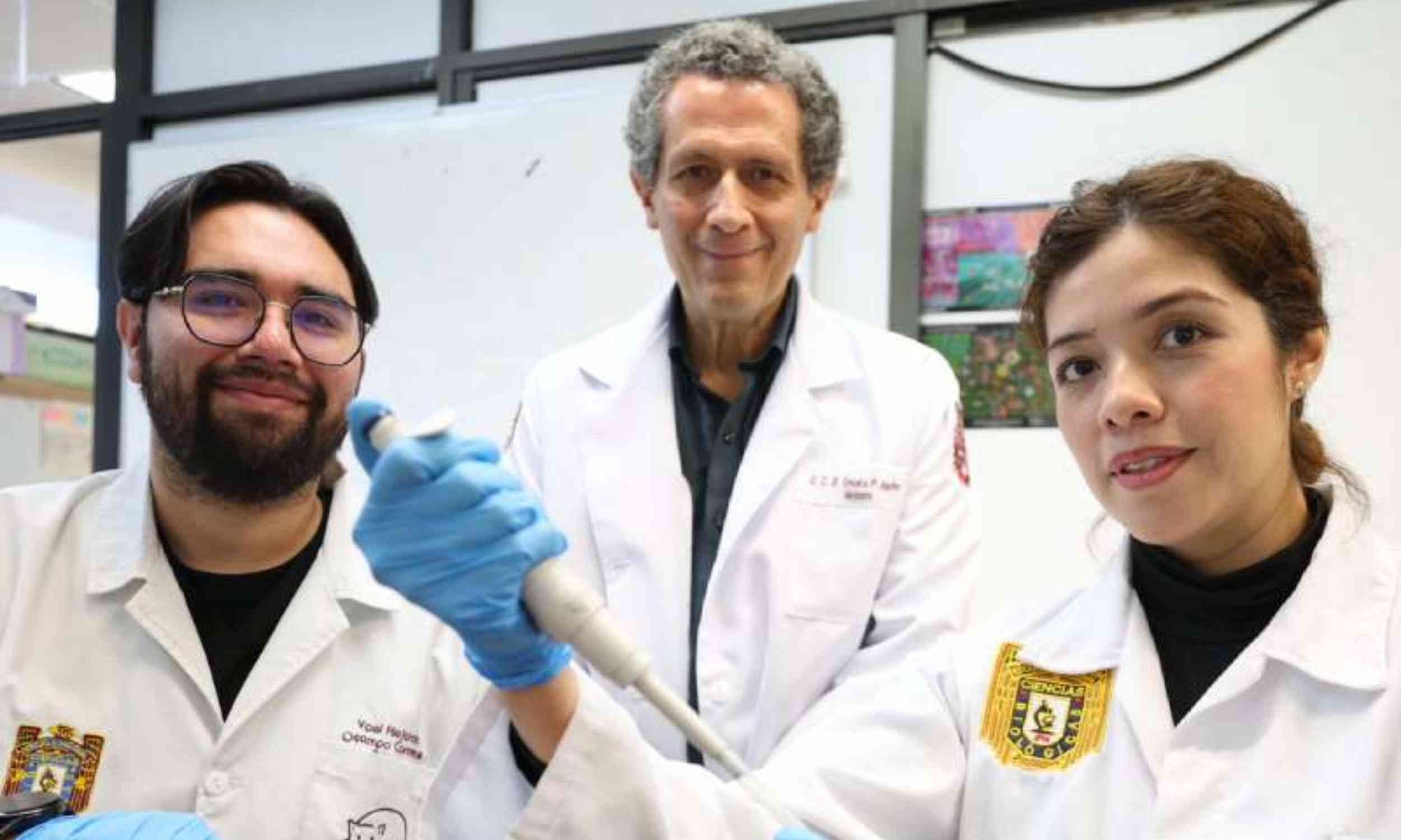
Researchers at the National Polytechnic Institute (IPN) led by Jorge Vela Ojeda have shown that bone marrow stem cells can be guided to grow into multiple tissue types.
The tissues did not emerge as a single uniform mass but instead developed into distinct forms that reflect the structure of bone, cartilage, muscle, and fat.
That distinction poses a new challenge, because shaping tissue in controlled conditions does not guarantee it will survive or integrate after implantation.
Multiple outcomes of stem cells
Mesenchymal stem cells sit at the center of the project, a marrow population that can become bone, cartilage, and fat under the right cues.
Unlike blood-forming stem cells, they are nonhematopoietic, meaning they belong to the system of tissue that supports bone marrow – not than the system that produces blood.
Researchers value them not solely for their potential, but also for the repair signals they release around damaged tissue.
That dual purpose of building and signaling explains why they keep appearing in repair studies.
Harvesting from original sources
The raw material in this study is derived from bone marrow, the soft tissue inside bones that houses several stem cell populations.
Vela said the simplest collection route is aspiration from the iliac crest, the upper rim of the pelvis, via a needle.
The amount of material found there naturally is small. However, the IPN group says the volume can be expanded in the laboratory before use.
That ability to multiply scarce cells yields a tiny sample into something large enough to test.
Scaffold structure support
Once expanded, the cells were placed on scaffolds, 3D-printed supports that give growing tissue a shape and a surface to grip.
Researchers then matched a construct to a stubborn fracture or another damaged site instead of growing an undirected mass.
The goal was to make bone, connective tissue, and muscle that fit a non-healing break or a specific organ.
Structure is not cosmetic here, because geometry can decide whether repaired tissue joins the body or fails under stress.
Healing signals at work
Repair does not depend only on the cells settling in and becoming permanent residents. Medical interest centers on the proteins and tiny vesicles these cells release. Observation focuses on which ones calm inflammation and help new blood vessels form.
This matters because a damaged site often needs a better healing environment before it can rebuild itself.
Even so, a construct that works in a dish can behave differently once blood flow, immune signals, and forces enter the picture.
The challenge of consistency
Before any implant reaches a patient, science has to survive a much less glamorous test: manufacturing discipline.
Cells kept in culture too long can mutate, drift into the wrong identity, or grow in ways no one intended.
Regulators look for sterility, purity, stable behavior, and evidence the product will not create new harm after implantation.
Those checkpoints slow the field down, but they also separate credible regenerative medicine from wishful marketing.
Guardrails for new therapies
International guidance is clear that complex cell products should not immediately transfer to promising laboratory results into routine care.
The International Society for Stem Cell Research (ISSCR) guidelines state that safety and effectiveness must be shown in clinical trials before standard use.
Long-term monitoring may also be necessary because transplanted cellular products can persist and create later problems.
Thus, the team’s next step – patient use with IMSS support – will be much more demanding and difficult.
Knowledge from the clinic
Experience also shapes how fast a project like this can mature. After 23 years of leading hematology at a specialty hospital in Mexico City, Vela has seen many failed laboratory results.
“It will help this field develop much faster,” said Ojeda.
That promise still depends on better experiments and clear evidence, not on automation alone.
Competing in regenerative medicine
Mexico has now entered the field because of this research, but Vela noted that the United States, Spain, England, and Germany have gained the most ground.
Regenerative medicine advances when biology, materials, surgery, and regulation see progress.
IPN’s result matters because it links a public university, a medical school, and a national health system around tissue that will not heal.
Whether that collaboration becomes a therapy will depend on reproducible results, not on how dramatic the early milestone sounds.
The hard part is no longer shaping bone-marrow cells into replacement tissue, but transporting that tissue safely to the clinic.
If researchers can bridge the gap with clean manufacturing, trials, and proven follow-up, regenerative medicine in Mexico has a bright future.
This study is published by the National Polytechnic Institute.
—–
Like what you read? Subscribe to our newsletter for engaging articles, exclusive content, and the latest updates.
Check us out on EarthSnap , a free app brought to you by Eric Ralls and Earth.com.
—–