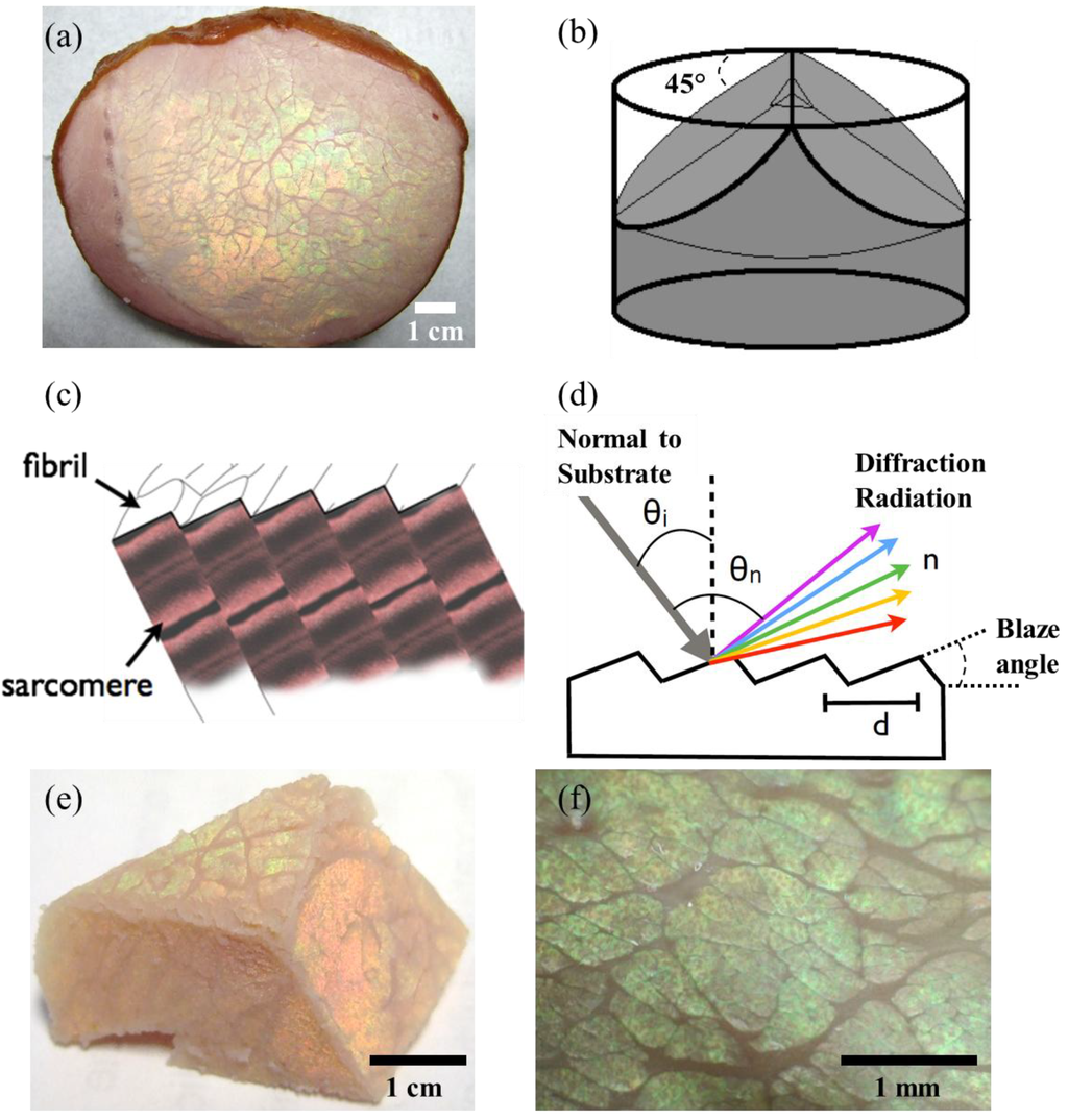
Iridescence is an optical phenomenon in which the light is diffracted from a photonic structure at a specific wavelength or a rainbow-like colour [ 1 ]. The iridescent colours are not caused by pigments, but by the interference of light with the morphology of the structure [ 2 3 ]. These photonic structures comprise of periodically ordered materials (or air gaps) with varying refractive index. The interference of light with the structure and its scatter causes a photonic effect in which only a dominant wavelength or multiple wavelengths are scattered back. Photonic structures including surface gratings, multilayers and thin films have been observed in nature [ 4 ] on the surface tissues or cuticle of a variety of species including birds [ 5 6 ], insects [ 7 ], marine molluscs [ 8 ] and plants [ 9 ]. The diversity in photonic structures might provide camouflage, warning colouration, superiority in reproductive behaviour, signal communication, thermoregulation and conspecific recognition. An analogous, but non-evolution-driven, photonic effect is also noticed in meat cuts, which display a rainbow-like multicoloured iridescence. Because it is difficult to distinguish the photonic effect from the original absorption colour of the meat, consumers may associate this phenomenon with spoilage, chemical or bacterial contamination, which may lead to refusal of the meat products. Considering the importance of this optical phenomenon in meat industry and costumer misconception, there is a need to investigate the factors causing meat iridescence to take preventive measures to minimise consumer concerns [ 10 ].
iliocostalis
Thunnus albacares
Muscular tissues are formed by elongated cells in a regular arrangement showing a well-ordered structure [ 11 ]. When viewed at specific angles, such photonic structures may produce iridescence. The orientation and spacing of the ordered structures are different from muscle to muscle and type of species; the iridescence, if present, is also likely to be different. For instance, different types of muscle tissues could produce different colours and intensities. It has been found that fish tissue slices produce colours that vary depending on the species and environmental conditions [ 12 ]. Muscle fibres in mammals are supported by structures formed by myofilament proteins grouped in fibrils that form cell fibres; when cross-sectioned at a certain angle, the diffracted light may produce iridescence depending on the structure. The arrangement of the cells in the whole muscle may have a different order and/or orientation depending on the muscle tissue type, however, it is usually homogenous. In fact, certain cuts of pork and beef exhibit iridescence [ 13 14 ], while others do not. In the case of beef, iridescence is most frequently found in semitendinosus muscle; it has a homogenous size distribution of cells and cylindrical fibrils that form a regular grid in transverse sections [ 15 ]. Several studies have investigated the association of iridescence with physiological factors such as pH, phosphate concentration, maturity, cooking temperature, fat thickness, surface roughness, shear force, animal type and sex type [ 16 17 ]. Along with these factors, iridescence in muscle tissue can be directly associated with water content. Since water is the main composition of the cell, changes in water content can cause evident changes in structure or optical properties [ 18 ]. Thus, the water content can be related to other factors like pH or ionic strength [ 19 ]. Therefore, characterisation of the structure and optical properties of the materials is crucial for understanding iridescence in natural structures. Previous studies have shown that iridescence in muscle tissue appears at a wavelength range seen as green, orange or red colours [ 20 ]. In recent investigations of cooked beef 21 ] and yellow fin tuna () [ 22 ], multilayer interference has been suggested as an explanation for the diffracted colours with strong angular dependence. In this work, we investigate the iridescence of cooked pork muscle tissue and report the intensity of the diffracted light in real time during controlled drying. We show that the intensity of iridescence is dependent on the water content of the tissue and the diffraction angle.