Due to its broad pharmacological effects, including anti-inflammatory and antioxidant properties, curcumin has been widely investigated in clinical studies. To overcome its low water solubility, poor bioavailability, and rapid metabolism, curcumin is formulated as nanocurcumin using different nanocarriers, such as liposomes, polymers, conjugates, cyclodextrins, micelles, dendrimers, and nanoparticles [ 293 ]. Transferrin-conjugated poly (lactic-glycolic acid) (PLGA) nanoparticles have been demonstrated to improve the bioavailability of curcumin to the brain and reduce Aβ deposition and tau hyperphosphorylation in the AD model [ 294 ]. Similarly, different formulations of nanoparticles have been shown to inhibit aggregation of Aβ and reduce depressive-like behavior and oxidative stress in AD models [ 295 296 ]. Intra-arterial administration of resveratrol (RES)-encapsulated nanoparticle (RES-NP) in a rat transient middle cerebral artery occlusion (t-MCAO) enhanced the resveratrol bioavailability and its brain-penetration, resulting in reduced infarct volume, and attenuated oxidative stress [ 297 ], brain edema, and neuronal apoptosis. The treatment also contributed to neurogenesis, leading to improved neurological recovery [ 298 ]. In a cerebral palsy rabbit model, intravenous treatment of dendrimer-based N-acetyl--cysteine (NAC) [ 299 ], a glutathione precursor with antioxidant and anti-inflammatory properties [ 300 ], reduced neuroinflammation and neurological injury, and improved motor function. In general, formulating antioxidants in nanocarriers has enhanced their efficacy due to better stability and/or improved transport to the CNS than free antioxidants [ 301 327 ]. Nanocurcumin has been evaluated as an add-on therapy to Riluzole in a pilot randomized clinical trial for safety and efficacy in ALS [ 272 ] and AD patients as dietary supplements [ 274 ]. In another study, solid–lipid curcumin showed significantly improved cognition and mood in healthy older population [ 273 ].
Edaravone-loaded ceria nanoparticles have demonstrated to cross the BBB via receptor-mediated transcytosis and protect the BBB [ 328 ]. In addition to the antioxidant property of ceria nanoparticles, edaravone provided its effect against oxidative stress in a stroke model [ 328 ]. Jin et al. demonstrated that the treatment with edaravone-encapsulated agonistic micelles caused rapid infarct volume reduction, prolonged survival, improved axonal remodeling, and reduced behavioral deficits than free edaravone-treated animals [ 329 ]. Wang et al. reviewed nanotechnology-based strategies for the treatment of ALS, including antioxidant agents [ 330 ]. Nanoparticle-loaded edaravone has been tested on the postoperative effects in patients with cerebral hemorrhage. The nanoparticle-loaded edaravone showed reduced edema as compared to free edaravone treated group, significantly improved neurological function, and reduced the production and release of interleukin and tumor necrosis factor, which was considered beneficial to protect healthy brain tissue and other organs, and conducive to the recovery and healing [ 275 ].
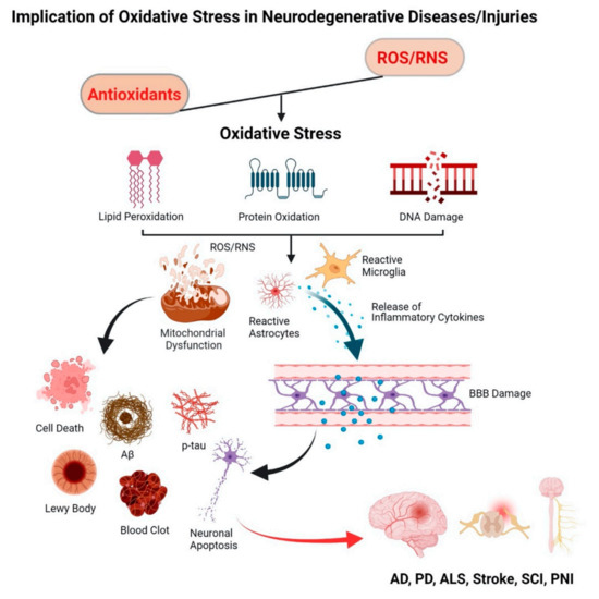
Antioxidant Enzymes
When they interact with free radicals, natural or synthetic antioxidants become inactive [ 331 ]. To continue to neutralize free radicals formed over a prolonged period, in chronic disease conditions, therapeutic levels of these antioxidants need to be maintained, which could be challenging, as repeated and high dosing cause dose-limiting toxicity in humans [ 331 ]. The main advantage of antioxidant enzymes is their catalytic mode of action [ 6 ]; hence, they can effectively neutralize free radicals at low doses. However, due to their short half-lives (5–11 min) [ 332 ], exogenously delivered antioxidant enzymes are ineffective in combating oxidative stress. Modifications such as PEGlylation and lecithinization improve their stability in the circulation [ 333 ] and fusion with cell membrane-penetrating peptides like a transactivator of transcription peptide or tetanus toxin fragment increases their ability to cross the BBB [ 334 ]. However, there are limitations to these modifications. Although PEGylated SOD (PEG-SOD) increases the enzyme’s stability in the circulation from 6 min to 36 h, PEG limits the permeability of the conjugated SOD across cerebral cell membranes [ 335 ]. Similarly, a chemical reaction involved in the fusion of different cell-penetrating or cell-specific peptides could cause denaturation and loss of enzyme activity [ 336 ]. In addition, the newly formulated hybrid enzyme could trigger immune-mediated anaphylactic responses to patients [ 337 ]. Intravenous delivery of SOD loaded into liposomes has shown to partially inhibit the infarct volume, but instability of liposomes in vivo (half-life ~4.2 h) limits the duration of SOD activity and, hence, its efficacy [ 338 339 ].
The recent effort includes formulations of antioxidant enzymes, SOD1, and catalase by electrostatic coupling of enzymes with cationic block copolymers called nanozymes [ 340 ]. In mice, this formulation demonstrated increased stability of enzymes in both blood and the brain and showed increased accumulation in the brain tissues than enzyme alone treated animals [ 340 ]. In a rat MCAO model, nanozymes reduced I/R-induced tissue injury and improved the sensorimotor functions [ 341 ]. In a moderate SCI rat model, treatment with nanozymes showed a recovery of locomotor functions, reduction of swelling, and post-traumatic cysts in the spinal cords of the treated animals [ 342 ]. Muzykantov’s group reviewed different nanocarriers to deliver antioxidant enzymes for vascular targeting in oxidative stress conditions associated with cardiovascular, pulmonary, and nervous systems [ 343 ].
2 O 2 -induced oxidative stress model in human neuronal cells and, subsequently, with the CAT-encapsulated nanoparticles (nano-CAT) in human astrocytes [ Our research group has been investigating the efficacy of antioxidant enzymes encapsulated in PLGA-based sustained release nanoparticles. The neuroprotective efficacy of the SOD-encapsulated nanoparticles (nano-SOD) was initially demonstrated in the H-induced oxidative stress model in human neuronal cells and, subsequently, with the CAT-encapsulated nanoparticles (nano-CAT) in human astrocytes [ 344 345 ]. In the MCAO model in rats, intracarotid administration of nano-SOD following 1 h of ischemia inhibited reperfusion injury. The treatment demonstrated improved neurological recovery and survival compared to controls (saline or SOD solution). There was evidence of neuronal recovery and regeneration with time in the above study [ 346 ]. The follow-up study in a thromboembolic rat stroke model, where tissue plasminogen activator (t-PA) was administered first for clot lysis followed by nano-SOD/CAT, both via the carotid artery, demonstrated the protective effect of the treatment. Significantly, the t-PA + nano-SOD combination treatment stimulated the migration of stem/progenitor cells from the subventricular zone and circulation, promoting neurogenesis. In contrast, this process was inhibited in the animals which received t-PA only treatment [ 347 ]. The above sequential treatment also inhibited edema formation, suggesting protection of the BBB from reperfusion injury [ 347 ]. In a separate study, we demonstrated aggravation of the BBB permeability when t-PA alone was administered via the carotid artery in the same thromboembolic rat stroke model [ 348 ]. Thus, the delivery of antioxidant enzyme nanoparticles in the above sequential treatment study protected the BBB from reperfusion injury and, also, from the effect of t-PA.